SMDEP Histology Laboratory Manual
Laboratory 1
#115 Gall Bladder, Hematoxylin & Eosin (H&E)
Open with WebViewer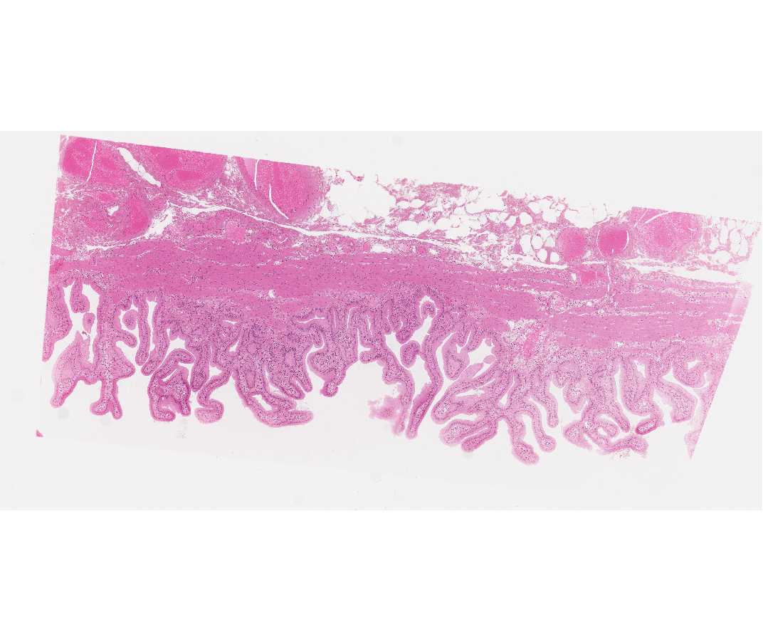
This is a portion of the gall bladder that has been stained with hematoxylin (blue) and eosin (red). Beginning with low magnification identify the nuclei of cells (blue) and their cytoplasm (light red). Go to higher magnifications to see these in more detail.
#107 Pancreas, Acid Fuchsin and Toluidine Blue
Open with WebViewer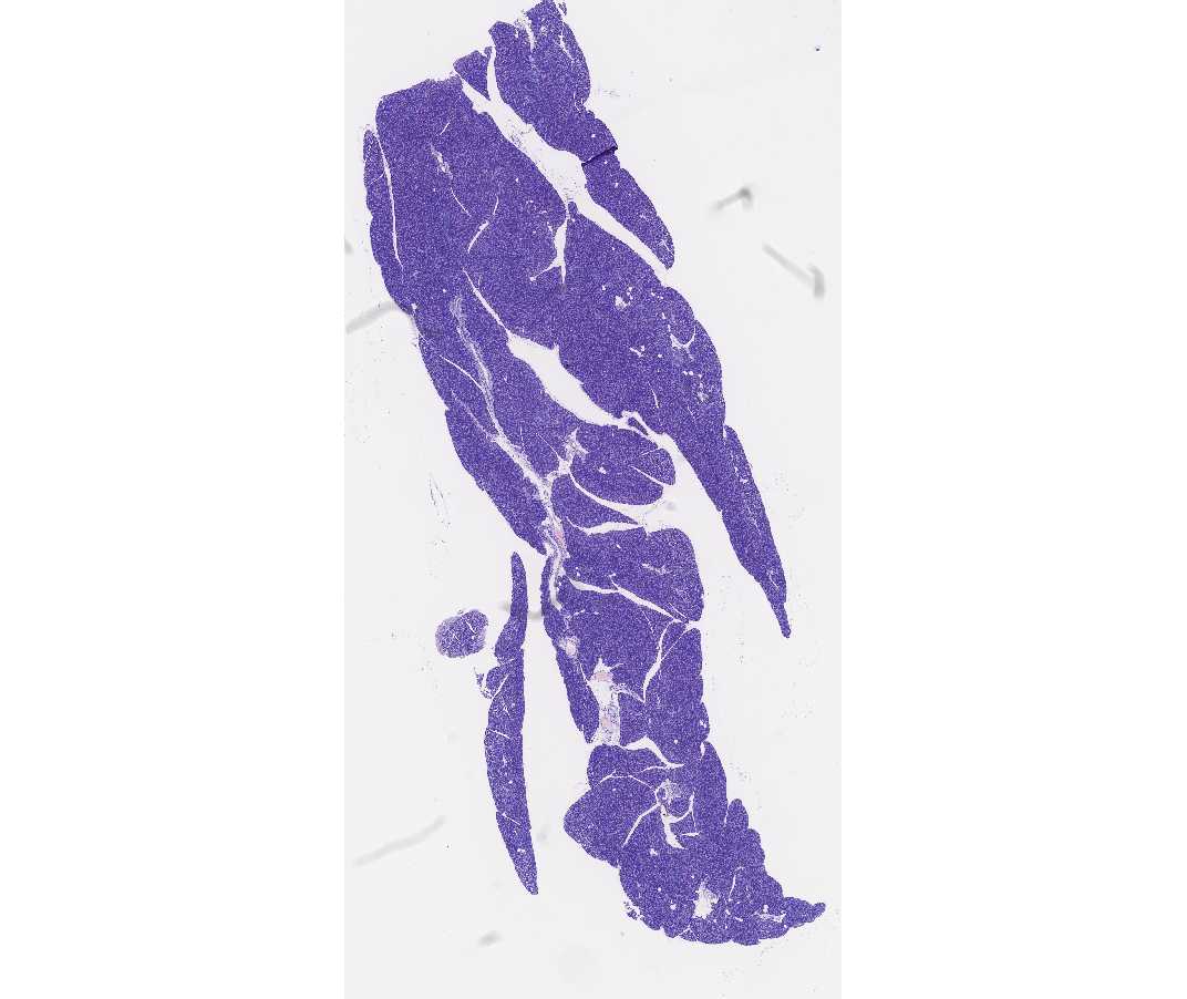
The cells of the pancreas store digestive enzymes in granules (pink) that are in the apical region. The basal region of these cells contains rough endoplasmic reticulum (blue) where the enzymes are made.
#103 Kidney, PAS & Hematoxylin
Open with WebViewer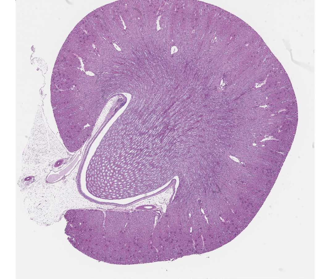
The kidney is a very complicated organ, but for this exercise you will be concentrating on the medullary region where there are many straight tubules running parallel with each other. Ask the instructors to help you find the medulla. There are longitudinal and cross sections of the tubules. Concentrate on the pale staining cuboidal cells that line the collecting tubules. The boundaries between the lining cells are easily visible.
#5 Trachea, H&E
Open with WebViewer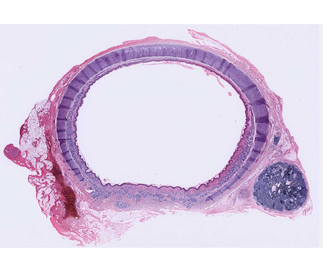
The lumen of trachea is lined by ciliated pseudostratified epithelium. The cilia appear as a pink fringe on the apical surface of epithelial cells.
#4 Thick Skin, H&E
Open with WebViewer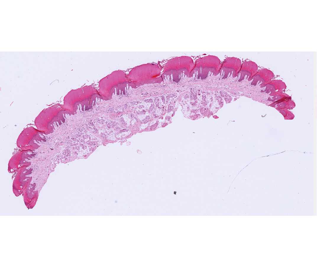
Examine the surface of the skin, noting that it is made up of a many layered epithelium. The superficial layers are dead cells with no nuclei.