SMDEP Histology Laboratory Manual
Laboratory 3
#32 Esophagus, H&E | |
| Open with WebViewer | |
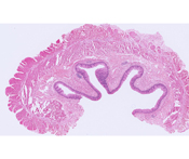 |
Examine the wall of the esophagus starting with the stratified squamous nonkeratinized epithelium, which covers the lumen. Underlying the epithelium is a layer of loose connective tissue and the muscularis mucosae. Note the thick muscularis externa. |
| |
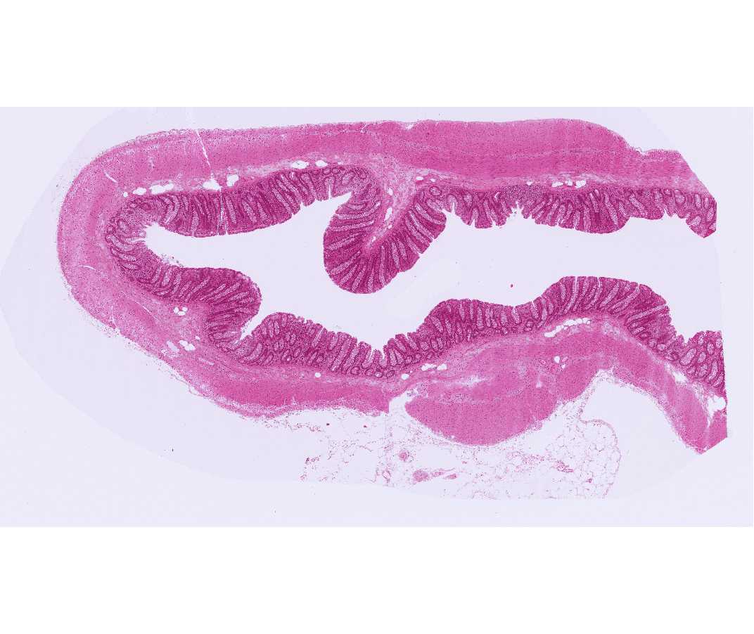
#117 Small intestine, H&E | |
| Open with WebViewer | |
 |
Identify the components of the wall of the small intestine. Use the laboratory manual as a guide to identify Paneth cells at the base of the intestinal crypts. These cells have accumulations of large acidophilic granules in their apical cytoplasm, and strongly basophilic basal cytoplasm. |
| |
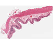
#39 Colon, H&E | |
| Open with WebViewer | |
 |
Note that the mucosa contains crypts and lacks villi. Also note the abundance of goblet cells in the lining of the crypts but not on the luminal surface; these features are diagnostic. |
| |
#106 Appendix, H&E | |
| Open with WebViewer | |
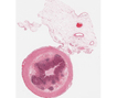 |
Examine the components of the wall of the appendix and compare them with the small and large intestine. In the mucosa of the appendix there is extensive accumulation of lymphocytes. |
| |