SMDEP Histology Laboratory Manual
Laboratory 2
#4 Thick skin, H&E | |
| Open with WebViewer | |
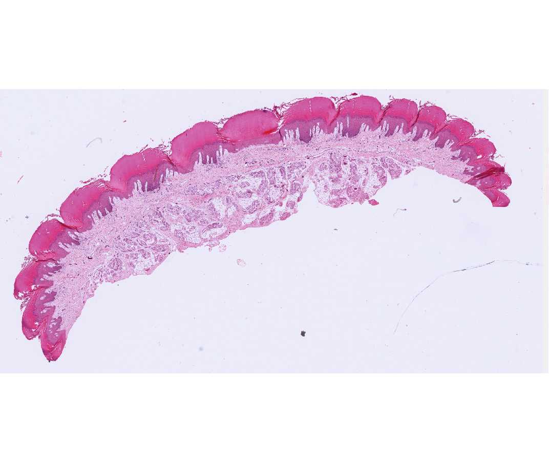 |
Below the epithelium (epidermis) is the dermis, which is dense connective tissue with many fibers and relatively few cells. Underneath the dermis is the hypodermis with a large amount of adipose tissue. |
| |
#12 Blood Smear, Wrights Stain | |
| Open with WebViewer | |
 |
The vast majority of cells are erythrocytes. Locate individual white blood cells and examine them at higher magnification. |
| |
#5 Trachea, H&E | |
| Open with WebViewer | |
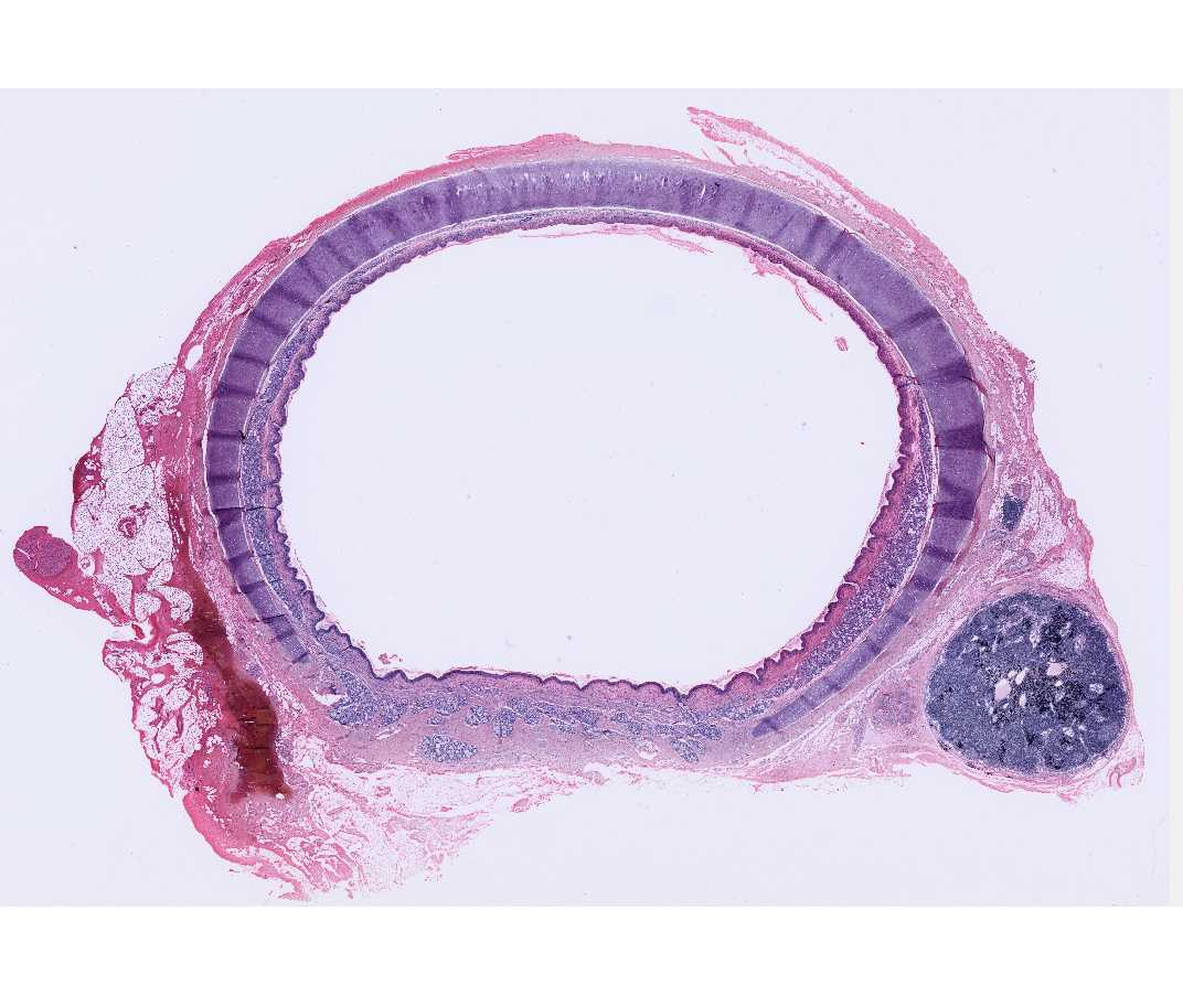 |
Find the ring of cartilage in the wall of the trachea which appears as somewhat shiny bluish tissue with many cells trapped in it. |
| |
#9 Dried Bone, Shaft of Tibia, Human, no stain | |
| Open with WebViewer | |
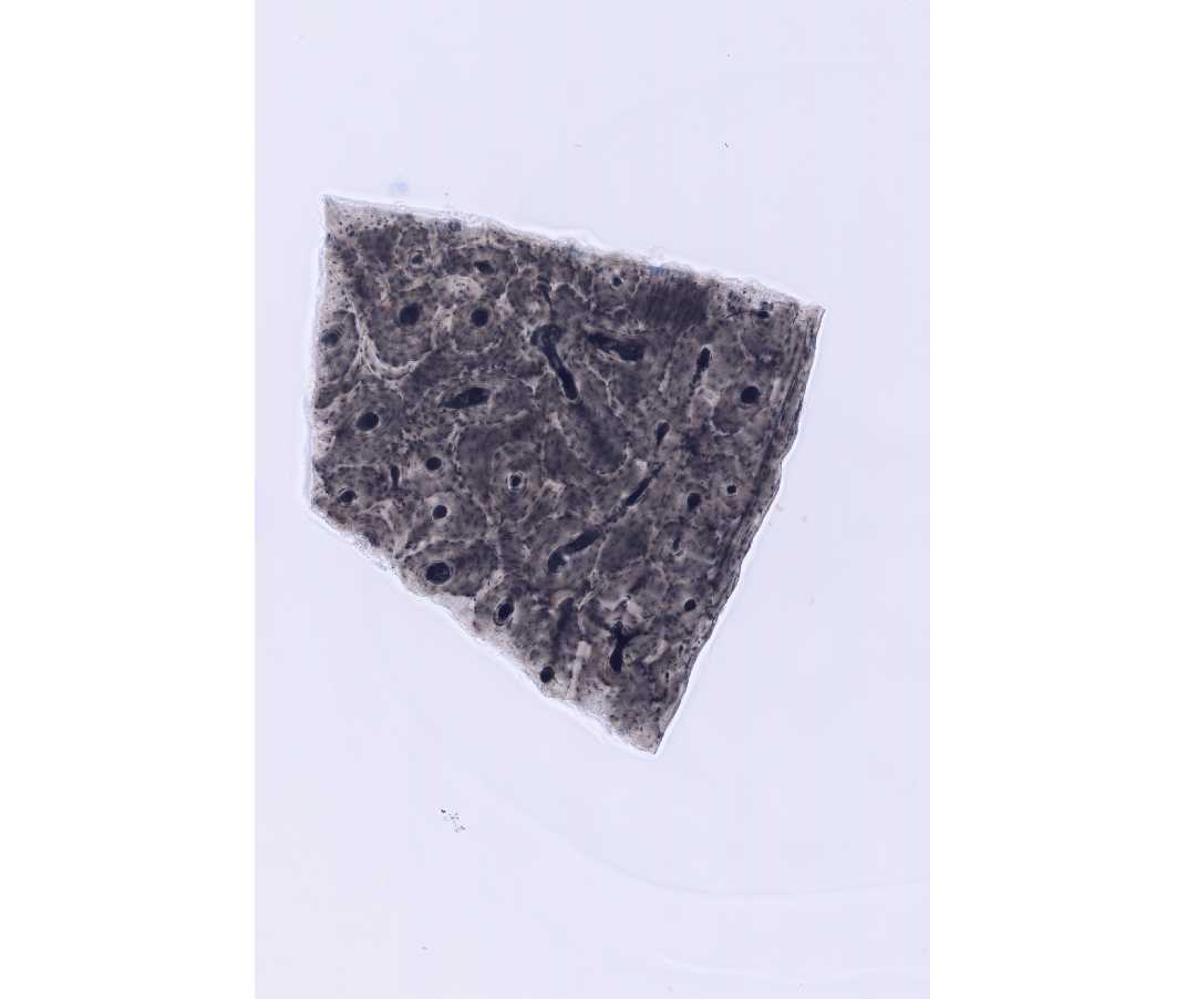 |
This section has been prepared by grinding down a piece of dried bone until it is thin enough to transmit light. The small dark regions are the sites where there were bone cells in life. There are also regions where blood vessels penetrate the bone. |
| |
#11 Rib, Cross Section, H&E | |
| Open with WebViewer | |
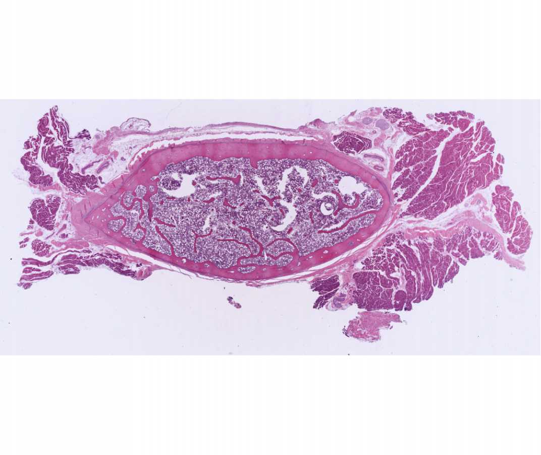 |
This section was prepared by decalcifying bone so that it could be cut and then staining it. There are some portions of surrounding muscle and connective tissue outside the bone and some marrow on the inside. |
| |
#116 Tongue Skeletal Muscle, H&E | |
| Open with WebViewer | |
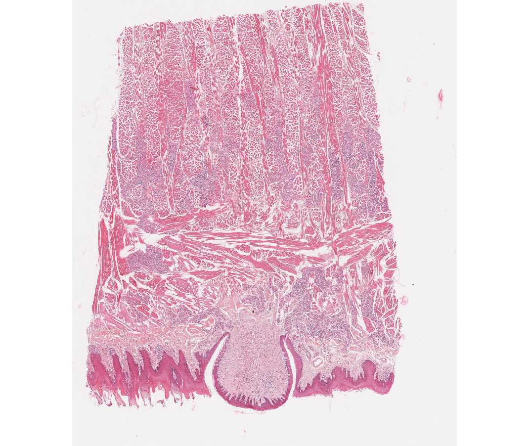 |
The outer surface of the tongue contains projections (the papillae). The skeletal muscle runs in various directions in deep regions. Use high enough magnification to see the striations. |
| |
#39 Colon, H&E | |
| Open with WebViewer | |
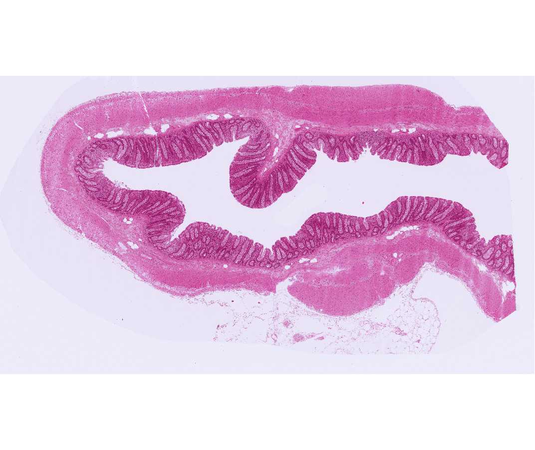 |
There are two bands of smooth muscle in the wall of the colon (and the other parts of the digestive tract). The cells in the outer layer are oriented parallel to the long axis of the colon; while cells in the layer interior to this are arranged circularly. At high magnification note that there are no striations and the nuclei are central. |
| |